A retinal hole is a serious condition that, if left unmonitored, can lead to permanent total or partial vision loss. Learn more about retinal holes, including their causes, risk factors, symptoms and treatment options here.
What is a retinal hole?
A retinal hole is a small tear in the macula. In medical literature, this condition is more commonly called a “macular hole” or “macular tear.” The macula is the center part of the retina, the very important tissue located at the back of the eye (also called the eye’s posterior segment). The retina is made up of light-sensitive tissue that allows people to see; when light hits the retina, it is converted into electrical signals that form our brain’s visual perception.
How common is a retinal hole?
7.8 out of 100,000 people, according to statistics from the advocacy group VisionAware. Age is the most common predictor of retinal holes, which typically occur in people over the age of 60, and a majority of patients are female.
What causes a retinal hole?
In short, a natural process gone wrong. As people age, fibers in the eye’s vitreous – the fluid that fills the eye and creates its globular shape – naturally shrink. However, if the shrinking fibers are too firmly attached to the retina, they can tear tissue as they retract, creating a retinal hole.
Is a retinal hole serious?
Retinal hole is a serious condition. The macula is the part of the eye that facilitates focused, centralized vision. This type of vision is important for reading, driving, and other tasks that require perception of fine detail. Accordingly, damage to this tissue can cause blurred, fuzzy, or otherwise distorted vision, especially in the middle part of the eye’s visual field.
Is a retinal hole an emergency?
Potentially. Depending on the retinal hole’s stage of progression, the condition might be considered a medical emergency. When the retinal hole reaches an advanced stage, it can cause retinal detachment, which can lead to permanent partial or total vision loss. However, it should be noted that even if the retinal hole hasn’t progressed to an “emergency” level, it is still highly serious and should be addressed with an eye doctor immediately.
What is retinal detachment?
Retinal detachment happens when the retina becomes detached from its underlying tissue. Retinal detachment is considered an emergency scenario, as it very rapidly leads to permanent partial or total vision loss.
What causes retinal detachment?
There are several different causes of retinal detachment. These causes include aging, injury, or changes due to other medical problems, such as diabetes or cancer. Retinal detachment can also be caused by other ocular conditions, such as age-related macular degeneration.
What are the symptoms of retinal detachment?
The sudden appearance of “floaters,” flashes, and reduced vision – according to the Mayo Clinic, these are the main symptoms to watch out for. Floaters are shapes that appear in a person’s visual field. They often appear as specks, strings, or cobwebs, and are caused by shadows created by the eye’s smaller anatomical structures.
How long can retinal detachment go untreated before permanent vision loss?
Not very long. Although the exact amount of time is highly dependent on individual and situational factors, retinal detachment typically leads to vision loss very quickly, and should be considered a medical emergency requiring immediate attention.
What are the risk factors for a retinal hole?
According to recent clinical research, primary risk factors include:
- Age: Incidence rates increase with age.
- Gender: Females have a higher rate of diagnosis than men.
- Race: Asian-Americans were found to have significantly increased risk.
- Other Eye Conditions: People diagnosed with cataract, aphakia, or pseudophakia had higher diagnosis rates.
How is a retinal hole diagnosed?
A retinal hole is diagnosed by an eye doctor. Your eye doctor may be an ophthalmologist (a medical doctor that is trained to perform ocular surgery) or an optometrist (a specially-trained doctor capable of providing non-surgical treatments). Most cases of retinal holes are discovered as part of diagnostic testing during routine medical examinations. This makes regular visits to your eye doctor extremely important, especially if you are over age 50.
How is a retinal hole treated?
A retinal hole is most commonly treated using traditional or laser surgery. Traditional surgery is performed using a method called vitrectomy, whereas laser surgery is performed using a laser that heats tissue in the back of the eye to “seal” holes in the macula.
- During a vitrectomy, an ophthalmologist removes the vitreous fluid pulling on the retina and injects a gas bubble to hold the eye’s physical structure in place. According to the American Society of Retina Specialists, vitrectomy has a success rate over 90%, and it is even possible for patients to recover some or all of their lost vision. Vitrectomy is considered an invasive option compared to other therapies.
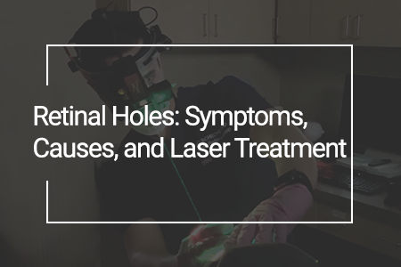
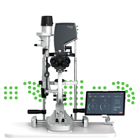
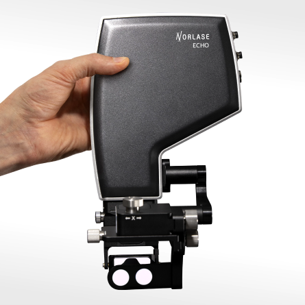
- During laser treatment, an ophthalmologist uses a laser device to perform “photocoagulation” of the tissue surrounding the hole. As the name implies, photocoagulation uses light (“photo”) to seal (“coagulate”) the tissue by heating it to a precise temperature. The heat triggers local cells to produce the proteins that create scar tissue, closing the holes.
It is worth noting that due to the costs and risks associated with any type of surgery, a doctor may opt to monitor very small holes over time instead of treating them immediately.
What are effective prevention strategies for retinal holes?
Unfortunately, there are no clinically proven ways to “prevent” retinal holes. Age, gender, comorbid conditions, and injury are generally considered uncontrollable factors. Systemic factors like diabetes can play a tangential role, so as always, it’s a good idea to eat well, exercise, and avoid stress wherever possible.
That said, routine visits to your eye doctor are crucial. By visiting your eye doctor regularly (at least twice annually, especially if you are over age 50), you can improve your chances of “catching” and diagnosing eye conditions, including retinal holes, as early as possible. Early detection and treatment is pivotal for achieving the best treatment outcomes possible, so be sure to make regular eye doctor visits a health priority.
Sources
- https://www.nei.nih.gov/learn-about-eye-health/eye-conditions-and-diseases/macular-hole
- https://visionaware.org/your-eye-condition/guide-to-eye-conditions/macular-hole/updates-in-macular-hole-treatments-and-recovery/macular-hole-statistics
- https://www.mayoclinic.org/diseases-conditions/retinal-detachment/symptoms-causes/syc-20351344
- https://www.nei.nih.gov/learn-about-eye-health/eye-conditions-and-diseases/retinal-detachment
- https://jamanetwork.com/journals/jamaophthalmology/fullarticle/2603482
- https://www.asrs.org/patients/retinal-diseases/4/macular-hole

DIDN’T FIND WHAT YOU’RE LOOKING FOR?
Let us know how we can help!
DIDN’T FIND WHAT YOU
WERE LOOKING FOR?
Let us know how we can help!


