What Is Retinopathy of Prematurity & How Common Is It?
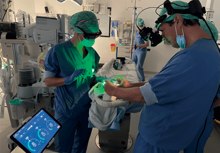
Dr. Frank T. Kerkhoff using the Laser Indirect Ophthalmoscope to perform laser surgery on an infant.
What Is Retinopathy of Prematurity & How Common Is It?
Dr. Frank T. Kerkhoff using the Laser Indirect Ophthalmoscope to perform laser surgery on an infant.
Retinopathy of prematurity is a disorder that affects thousands of premature infants each year. Learn more about the disorder, signs, stages of severity, treatment options and more here.
What is retinopathy of prematurity (ROP)?
Retinopathy of prematurity (ROP) is an eye disorder that mainly affects premature infants. Smaller infants, as well as infants born especially early, have a higher chance of being born with the disorder. Unlike many significant ocular conditions, ROP usually develops in both eyes at the same time. According to the National Eye Institute, ROP is “one of the most common causes of visual loss in childhood and can lead to lifelong vision loss and impairment and blindness.”
How common is retinopathy of prematurity?
14,000-16,000 babies are born with retinopathy of prematurity each year, according to the advocacy group March of Dimes.
Is retinopathy of prematurity present at birth?
Retinopathy of prematurity is present at birth, although the abnormalities that lead to the condition develop earlier in the gestation process.
How does retinopathy of prematurity affect vision?
Retinopathy of prematurity occurs when blood vessels in the eye grow in an abnormal way. If the extent of this abnormal growth is severe enough, the child may suffer retinal detachment, a severe condition that can lead to permanent total or partial vision loss.
Is retinopathy of prematurity degenerative?
Retinopathy of prematurity is potentially degenerative. If left untreated, it can progress over a five-stage sequence characterized by increasingly abnormal blood vessel growth and medical consequences, such as visual distortion or vision loss. However, some cases of ROP do not progress. In these instances, blood vessel growth normalizes over the course of an infant’s development. For this reason, early-stage ROP is often monitored closely by physicians to determine if treatment is necessary.
Is ROP curable?
Medical intervention can help resolve the causes and symptoms of retinopathy of prematurity. Often, intervention will slow or prevent visual changes or vision loss, but there is no guarantee that treatment will provide full relief or remediation. Early-stage disease sometimes resolves on its own, so physicians will often only treat patients that display clear signs of advanced, aggressive disease – Stage III-V on the chart included below.
What causes retinopathy of prematurity?
The root “cause” of retinopathy of prematurity is complicated. As an infant gestates in the womb, blood vessels in the eye generally grow along a normal, healthy trajectory. However, when an infant is born prematurely, a lack of oxygen in certain parts of the eye may cause the blood vessels to grow along an abnormal path, leading to the anatomical condition and consequences that characterize the disorder.
What are the stages of ROP?
There are five stages of retinopathy of prematurity, classified in increasing order of severity. When an infant is observed to be in Phase III or above, treatment is considered. Severity is calculated by factoring:
- the severity of blood vessel abnormality
- the area of the retina where abnormal vessels are found
- the extent of vessel appearance
- whether the blood vessels are dilated or “torturous” (referred to as “plus” disease; a sign of advancing disease progression, and often a trigger for intervention)
- whether the condition is apparent and aggressive in the back-most part of the eye
These factors are assessed to categorize the condition as being in one of the following stages (as defined by the NEI):
- Stage I: Stage I ROP is characterized by “mildly” abnormal blood vessel growth. Many infants with this diagnosis do not require treatment, as the disorder and vision normalizes over the course of development. Along with Stage II, a majority of infants are diagnosed at this stage.
- Stage II: Stage II ROP is characterized by “moderately” abnormal blood vessel growth. Although this is more advanced than Stage 1, again, many infants with this diagnosis do not require treatment, as the disorder and vision normalizes over the course of development. Along with Stage I, a majority of infants are diagnosed at this stage.
- Stage III: Stage III ROP is characterized by “severely” abnormal blood vessel growth. At this point, the blood vessels are diverted from their normal growth path. Some infants with Stage III ROP will eventually develop normal vision, but patients with “Plus” disease (as mentioned above) are typically flagged for treatment. Intervention at this stage can help prevent retinal detachment.
- Stage IV: Stage IV ROP indicates a partially-detached retina. This is the stage at which consequences become very acute and future vision loss highly likely.
- Stage V: Stage V ROP indicates complete retinal detachment. This stage is characterized by severe, if not total, vision loss.
In recent years, scientists have trialed the use of Artificial Intelligence capable of “reading” clinical photos to more accurately categorize disease severity using the 5-stage system.
Testing and diagnosis
Testing for retinopathy of prematurity occurs when a baby is born prematurely. Because there are no reported symptoms, protocol-based testing is used to look for signs of the disorder. This testing is done using a standard eye exam, which typically occurs around the 4 week mark post-birth.
How is ROP treated?
Retinopathy of prematurity is most often treated using laser therapy or cryotherapy. These treatment methods are especially preferable for treating earlier stages. Unfortunately, given the nature of the disorder, treatment involves a “tradeoff” whereby the treating physician seeks to save central vision at the potential cost of peripheral vision.
- Laser therapy involves the use of a laser photocoagulator to burn away blood vessels in the eye’s periphery. Because peripheral vessels are not essential to sight, cauterizing this tissue can block the inward spread of abnormal growth, preserving central vision and saving sight.
- Cryotherapy takes a similar approach: however, instead of peripheral vessels being burned away, they are frozen to the point of cell death.
If the condition progresses to the point of partial or total retinal detachment, more traditional surgical interventions, such as vitrectomy or a scleral buckle, are used.
Relevant clinical research on ROP
The diagnosis and treatment of retinopathy is a primary area of concern for ophthalmic research. Because early detection, monitoring, and intervention (when necessary) is crucial to ensuring positive life outcomes for infants around the world, scientists are exploring new ways to catch and manage the disease as early as possible. For more information, please visit:
Visit Norlase to learn how we are changing the way doctors treat. Check out our blog for more interesting topics related to ophthalmic lasers.
Sources
- https://www.nei.nih.gov/learn-about-eye-health/eye-conditions-and-diseases/retinopathy-prematurity
- https://www.marchofdimes.org/complications/retinopathy-of-prematurity.aspx#:~:text=About%2014%2C000%20to%2016%2C000%20babies,become%20legally%20blind%20from%20ROP
- https://eandv.biomedcentral.com/articles/10.1186/s40662-020-00206-2
- https://www.ncbi.nlm.nih.gov/pmc/articles/PMC5806220/

DIDN’T FIND WHAT YOU’RE LOOKING FOR?
Let us know how we can help!
DIDN’T FIND WHAT YOU
WERE LOOKING FOR?
Let us know how we can help!

